Effect of inspiration degree on the measurement of lung nodule volume
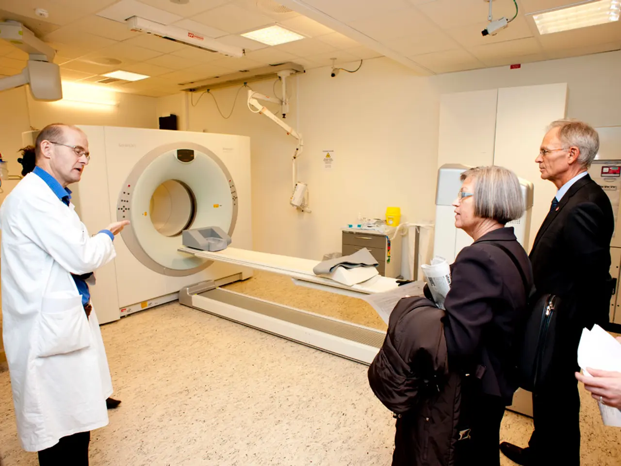
In a recent study conducted at a French university hospital in 2022, researchers investigated the effect of inspiratory level on lung nodule volumetry in CT scans. The study included 403 examinations of patients who had one or more nodules, as defined by the Fleischner Society.
The primary objective of the study was to explore the impact of inspiratory level on nodule volumetry. The researchers found that the inspiratory level during CT scanning significantly affects the reliability of lung nodule volumetry. This is primarily because variations in lung volume impact nodule size and shape as captured by imaging.
The study revealed that the amplitude of respiratory effort influences lung inflation, causing nodules to appear larger or smaller depending on the phase of respiration, thereby affecting measured volumes. Nodule-specific characteristics, such as nodule type (solid, part-solid, ground-glass), shape, and location, also predict how much volume measurements vary with respiratory changes. For example, nodules attached to pleura or within fissures may show less volume variability compared to those suspended in lung parenchyma.
To mitigate these variations, the researchers emphasised the importance of standardising the inspiratory volume during CT acquisition. They found that instructing patients to hold breath at full inspiration helps minimise these variations and improve volumetric measurement consistency.
The study also found that the volumetry technique itself benefits from controlling inspiratory levels to reduce errors in quantitative analysis of nodule volume over time or between scans.
The researchers used Vitrea's "CT Lung Nodule Analysis" software for semi-automated segmentation of lung nodules and lung parenchyma. Manual editing was done if necessary by the operator. For each included nodule, its size, morphology, lobar topography, closeness to the pleura, and the regularity of its margins were listed.
The study excluded nodules that were typical and atypical perifissural, calcified, excavated, or from technically insufficient/unconform exams. The nodules had a greater diameter (on inspiratory CT-scan measured in native axial plane) of at least 6 mm.
The examinations were consulted by a single operator, a radiologist with 4 years of training in CT. CT scans were acquired on 320-row scanners with specific technical parameters. The nodule and lung volumes variations between inspiratory and expiratory acquisitions were calculated for each pair of exams using the formula: volumevariation(%)=VBMI−VRVBMI×100.
The secondary objective of the study was to query the amplitude of respiratory effort and nodule-specific characteristics as predictive factors of nodule volume variations. While explicit data linking inspiratory level and variability in volume measurements is often studied in the context of nodules’ radiomic and morphological features, predictive modeling incorporating factors like nodule size, mean CT value, and solid component (CTR) has shown improvement in assessing nodule behaviour and malignancy risk, indirectly reflecting the impact of these volumetric accuracy factors.
In summary, higher and more uniform inspiratory levels aid in reliable lung nodule volumetry on CT by reducing respiratory-induced variability. The amplitude of respiratory effort and individual nodule features serve as key predictors of volumetric variation. This consistency is critical for accurate longitudinal assessment of nodules for malignancy risk and treatment monitoring.
Raw-image data was processed using iterative reconstruction (AID3RD) or deep-learning reconstruction (AiCE). The study used a monocentric retrospective observational design.
In the field of medical-conditions, the study highlighted that the inspiratory level during health-and-wellness checks like CT scans significantly affects the reliability of lung nodule volumetry, as variations in lung volume can impact nodule size and shape. The study further pointed out that predicting nodule volume variation also depends on nodule-specific characteristics, such as nodule type, shape, and location, emphasizing the importance of standardizing the inspiratory volume during CT acquisition for more accurate health analysis.


