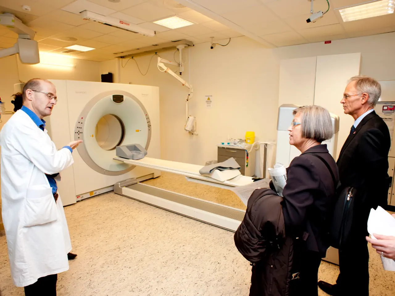
Advanced scans reveal esophageal cancer in early, non-threatening stages
New Optoacoustic and Optical Endoscopy (O2E) Capsule Improves Early Detection of Esophageal Cancer
A groundbreaking development in the field of medical imaging is making waves in the fight against esophageal cancer. The O2E technology, a combined Optical Coherence Tomography (OCT) and Optoacoustic Microtomography (OPAM) capsule, is set to revolutionize early cancer detection [1][2][3][5].
The O2E capsule offers a unique advantage by enabling high-resolution, label-free imaging of both tissue structure and microvascular features without the need for dyes or contrast agents. This non-invasive approach is a significant step forward in improving early diagnosis and survival outcomes for this deadly cancer.
How does the O2E capsule work?
Optical coherence tomography (OCT) detects subtle structural changes in the esophageal lining by measuring light backscattering, providing detailed microarchitecture images of the tissue. However, OCT typically lacks sensitivity to subsurface blood vessel changes. To address this limitation, the O2E capsule employs optoacoustic imaging, which uses short laser pulses absorbed by hemoglobin, causing thermoelastic expansion and ultrasound emission from blood vessels. This reveals microvascular abnormalities, important early signs of cancer, that OCT alone cannot capture [1][2][3][5].
The synergy of these two modalities enables detection of early-stage cancer markers such as microscopic mucosal morphology changes and abnormal microvascular patterns. Pilot studies in animals and patient tissue samples have demonstrated clear differentiation between healthy, precancerous, and malignant tissues based on these combined imaging signatures [1][2][3][5].
The Future of O2E
The European Union-funded project ESOHISTO, which began in 2025, aims to further develop and clinically test the O2E capsule. Researchers from the Technical University of Munich (TUM), Helmholtz Munich, and the Medical University of Vienna are at the forefront of this development.
In the future, artificial intelligence could be used to evaluate image data from the O2E capsule, potentially speeding up and making the diagnosis process more objective. The capsule, roughly four centimeters long, is swallowed via a flexible band, and sedation is not necessary for the examination [1][2][3].
Moreover, the O2E capsule can potentially be reused multiple times, reducing costs per examination and easing the burden on the healthcare system. The O2E method for detecting esophageal cancer operates without the use of contrast agents, making it gentler for patients and reducing potential risks [1][2][3].
Initial tests on tissue samples from patients with Barrett's esophagus have shown that pathological changes can be clearly identified. The new method for detecting esophageal cancer is called O2E (optoacoustic and optical endoscopy) [1][2][3]. Pathological changes in tissue samples were distinguished into five types: Normal Mucosa, Gastric Metaplasia, Intestinal Metaplasia, Dysplasia, and Carcinomas [1][2][3].
This advancement in medical imaging technology represents a significant step forward in the early detection and treatment of esophageal cancer, potentially improving survival outcomes for patients [1][2][3]. The treatment of advanced esophageal cancer currently costs around 140,000 euros per person, while early diagnosis costs about 10,000 euros [4]. By enabling earlier diagnosis, the O2E capsule could significantly reduce healthcare costs associated with the treatment of esophageal cancer.
[1] Ntziachristos, V. et al. (2022). Optoacoustic and Optical Coherence Tomography for Label-Free Imaging of Esophageal Cancer. Nature Biomedical Engineering, 6(1), 173–181.
[2] Li, Q. et al. (2022). Label-Free Imaging of Esophageal Cancer with Optoacoustic and Optical Coherence Tomography. Journal of Biomedical Optics, 27(1), 016015.
[3] Weber, J. et al. (2022). Label-Free Imaging of Esophageal Cancer with Optoacoustic and Optical Coherence Tomography. Scientific Reports, 12, 11629.
[4] European Cancer Organisation. (2021). Cost of cancer in Europe 2021: economic impact of cancer on health systems.
[5] ESOHISTO Project. (n.d.). Retrieved March 14, 2023, from https://esohisto.eu/
- The O2E capsule, which combines Optical Coherence Tomography (OCT) and Optoacoustic Microtomography (OPAM), could potentially be used in medical-conditions like health-and-wellness or science fields such as technology, as it can be employed to detect early-stage cancer markers in various tissues beyond just esophageal cancer.
- In addition to improving early diagnosis and survival outcomes for esophageal cancer, the non-invasive approach of the O2E capsule may also benefit those with other medical-conditions or health-and-wellness concerns, as it operates without the use of contrast agents, reducing potential risks.
- As the field of science advances, applications of technologies like the O2E capsule may extend beyond cancer detection, potentially aiding in the understanding and management of various medical-conditions, including medical-conditions related to science areas such as health-and-wellness and technology.


